Side
View Technology
The novel Side View Technology, which was developed in Dr.
Chi-Hung Lin and Szecheng Lo's lab, allows us to observe
the cell grown on substrates from their sides with high spatial
resolution and in REAL TIME!
Using
this technology, you can easily observe the sides of cultured
cells under conventional optical microscopes. This technique
may be used in conjunction with virtually all other microscopy
techniques including bright field, dark field, phase contrast,
differential interference contrast (DIC), and fluorescence
microscopy. In contrast to confocal scanning microscopes,
by which the observed plane has to be scanned resulting in
largely decreased temporal resolution, our new technique bears
the ability to image the cell from its side in REAL TIME.
Owing
to the special optical configuration of the Side View Technology,
the spatial resolution can also tuned up to optical limits,
without the limitations of the mechanical components, such
as stepper motors. Consequently many fast and subtle biological
phenomena can be resolved. Moreover, cells or tissues at the
side view configuration could also be manipulated by optical
tweezers simultaneously. This totally novel combination
of observing and manipulating cells may open a new window
of biomedical research.
|

|
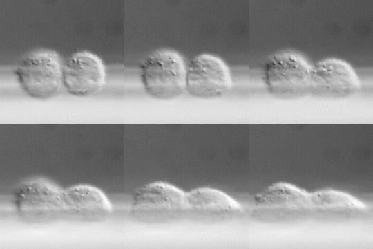
Cell
spreading observed by the Side View Technology. Two
Chinese Hamster Ovary (CHO) cells expressing integrin
aIIbb3
were put on a substrate coated with rhodostomin,
and the process of cell spreading was observed using
the Side View Technology with time-lapse recording.
The cells were shown to spread and flatten within about
1.5 hours. Similar technique was successfully applied
to platelets to observe their
cellular activities and spreading.
|
|

|
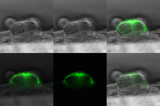
Cell
membrane glycoproteins decorated by a fluorescein-conjugated
lectin, concanavalin A (Con A) observed by the Side
View Technology. Con A recognizes a-linked
mannose, which commonly occurs in glycoproteins. As
shown here, the Side View Technology is capable of observing
cells with fluorescent microscopy.
|
|

|
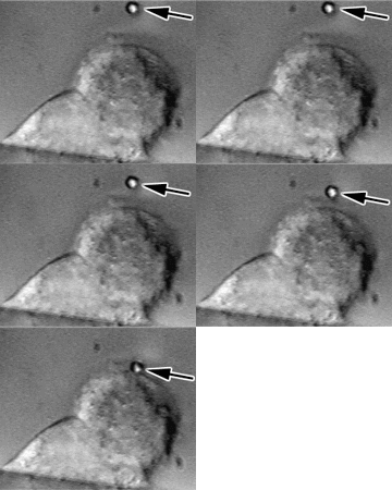
The
combination of side view microscopy and optical
tweezers. A bead coated with poly-L-lysine (arrow)
was trapped by optical tweezers
and put on the surface of a Chinese Hamster Ovary (CHO)
cell. The entire process was observed via the Side View
Technology. This combined technology can be used in
the observation of many cellular activities such as
endocytosis and intracellular signaling.
|
Links:
Updated
6/13/2013. Copyright© 2001
Jin-Wu Tsai. All rights reserved.
|
