Cellular
Mechanics Using Optical Tweezers
Microscopic
objects, including biological materials, could be remotely
manipulated with tightly focused beams of laser light (Ashkin
and Dziedzic, 1987). Focusing by high numerical aperture (N.A.)
objectives, the light pressure and optical gradient forces
could be used to hold, and therefore move, sub-micrometer
sized objects, even in the interior of cells (Ashkin et al.,
1990). In addition to micromanipulation, optical tweezers
could also be employed to biological force measurement (for
review, see Ghislain et al., 1994). Typically, the optical
tweezers using wavelengths that are less absorptive (and therefore
less destructive) for biological materials (700-1100 nm) could
easily exert pN forces.
In
our study, we have constructed an optical tweezers module
that is well-integrated with an inverted light microscope
for micro-manipulation and/or force measurements. Significant
progress has been made to improve both the sensitivity and
accuracy of the quantification procedures using the award
winning forward-scattered light analysis. We have also successfully
applied this new tool to investigate the molecular interactions
between an integrin aIIbb3
and a disintegrin (the snake venom, rhodostomin,
from Malayan pit viper, Calloselasma rhodostoma).
|

|
Cell
contact by optical tweezers. A HEK-293T cell was manipulated
to make contact with another cell already attached to
the substrate. Our results showed that the interactions
of cells began in a very short time ranging from tens
of seconds to several minutes, which suggested the need
of optical tweezers in the studies of cell-cell interactions
and the underlying molecular interactions.
|
|

|
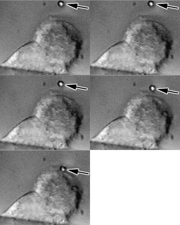
The
combination of side view microscopy and optical tweezers.
A bead coated with poly-L-lysine (arrow) was trapped
by optical tweezers and put on the surface of a Chinese
Hamster Ovary (CHO) cell. The entire process was observed
via the Side View Technology.
This combined technology can be used in the observation
of many cellular activities such as endocytosis and
intracellular signaling.
|
Further
Readings:
- Hsieh
CF, Chang BJ, Pai CH, Chen HY, Tsai JW, Yi YH, Chiang YT,
Wang DW, Chi S, Hsu L & Lin CH (2006) Stepped changes
of monovalent ligand-binding force during ligand-induced
clustering of integrin aIIbb3.
J
Biol Chem
281, 25466-25474.
- Hsieh
CF, Chang BJ, Pai CH, Chen HY, Chi S, Hsu L, Tsai JW &
Chi-Hung Lin (2004) Identification of stepped changes of
binding affinity during interactions between the disintegrin
rhodostomin and integrin aIIbb3
in living cells using optical tweezers. Proc
SPIE
5514, 215-224.
- Jin-Wu
Tsai. Using Optical Tweezers in Biological Force Measurements.
Master Thesis, National Yang-Ming University (2000).
Links:
Updated
6/13/2013. Copyright© 2001 Jin-Wu
Tsai. All rights reserved.
|
