細胞肝臟學
Cellular Hepatology
In
the studies of cellular physiology of hepatocytes (liver cells),
we mainly focus on the hepatobiliary secretory activities
and protein sorting/targeting mechanisms. We utilize several
well-differentiated hepatic cell lines that develop bile canalicular
(BC) structures in culture as our cell models. By applying
various fluorescent molecules as secretory markers, we are
able to observe their transportation/secretion processes by
confocal microscope. The movies shown below are examples of
2 different patterns of the secreted markers.
|
|
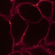
Secretion
of a fluorescent phospholipid analog, 1,2-dimyristoyl-sn-glycero-3-phosphoethanolamine
(DMPE), by the hepatoma cell line, HepG2. The fluorescent
molecules were incorporated into the basolateral membrane
of cells immediately after being added into the culture
medium, whereas the bile canalicular membrane showed
very faint signal, exhibiting a discontinuous hollow
among neighboring cells. The intensity of the fluorescence
signal in bile canaliculi (BC) then increased as time
goes on, indicating the lipid molecules are transported
to the BC domain. (Collaborated with Pei-Ling Lee in
Dr.
Chi-Hung Lin's lab.)
|
|
|
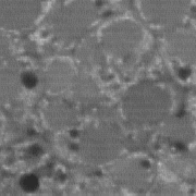
Secretion
of a fluorescent organic anion, fluorescein, by HepG2.
Fluorescein diacetate (FDA) was added into the culture
medium and diffused through the cell membrane. In cells,
FDA is then hydrolyzed by ubiquitous intracellular easterases,
which subsequently produced the fluorescent anion, fluorescein.
The process of the transportation can be visualized
by observing the fluorescence signal within BC lumen
(dark hollows). As the demonstration shown here, only
a subset of BC is capable of the transportation of fluorescein.
We found that the transportation ability of BCs is correlated
with certain protein sorting mechanisms in liver cells
(Lian et al., 1999).
|
Further
readings:
Links:
|